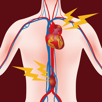
Abdominal aortic aneurysm
Definition
 The aorta is the largest vessel in the human body: starting from the heart, it first has a thoracic route (whose branches bring blood to the brain) and then an abdominal route (whose ramifications supply blood to the brain). intestine, liver, splanchnic organs, kidneys and lower limbs).
What is an abdominal aortic aneurysm?
The aortic aneurysm is a permanent dilation of the artery that affects the entire thickness of the wall. It can develop throughout its course, although the segment most frequently affected is the abdominal segment - explains Dr. outlet -. Another site frequently affected is the thoracic aorta and the abdominal aorta in the section where the visceral vessels are emitted”.
The aneurysm affects men more than women because they are more prone to arteriosclerosis. The latter being a typical disease of aging, it is easier to observe after the age of 65.
If, once found, it is not already large enough to be operated on (greater than 50 mm), it must be monitored over time using a non-invasive test such as the Doppler Ecocolor.
Causes
In 90% of cases, the aortic aneurysm is due to arteriosclerosis, a multifactorial disease caused by:
Familiarity.
Ageing.
Hypercholesterolemia.
Smoking.
Hypertension.
Diabetes.
Dyslipidemia.
Other factors that have not been studied as yet (infectious or chronic inflammation of the artery).
How does an abdominal aortic aneurysm manifest?
Abdominal aortic aneurysm is usually asymptomatic and is discovered, mostly, occasionally, by routine examinations (eg, abdominal ultrasound) done for other diagnostic questions.
When the aneurysm is about to rupture, the subject may feel very intense abdominal pain, often extending to the back, which simulates left renal colic.
In about half of cases of abdominal aortic aneurysm rupture, due to massive internal bleeding with hypovolemic shock, difficult to resolve. In the other half of cases, however, there is loss of blood, with the pressure falling and containment of the hemorrhage allowing emergency surgery before the actual rupture of the aorta occurs.
How is an abdominal aortic aneurysm diagnosed?
The first level examination that anyone can undergo for the diagnosis of an abdominal aortic aneurysm is ultrasound or, better still, Ecocolor Doppler which allows the abdominal aorta to be visualized in detail - continues the expert - . It is a non-invasive test, without contrast products and without the administration of ionizing radiation, indicated to assess the flow inside each vessel and the diameter of the aorta.
The dilated aorta can be:
Fusiform, more common, when the expansion is uniform and covers 360 degrees of the wall
Saccular, when it affects only one side of the aorta.
The natural history of abdominal aortic aneurysm is its gradual dilatation without symptoms until the time of rupture.
Surgical treatment
The treatment of self-limiting abdominal aortic aneurysm is exclusively surgical and can be:
Traditional (open): consists of replacing the dilated aorta segment and its reconstruction by the implantation of a plastic tissue prosthesis which can be straight (straight prosthesis) or Y-shaped (bifurcated prosthesis).
Endovascular: allows to treat the same pathology by placing a prosthesis from the inside the diseased aorta, accessing it through the femoral arteries through small holes. Using the angiograph, an angiography with contrast medium is performed and the aneurysm is observed, its relationship with the other arteries, then the type of prosthesis to be used is decided.
While traditional surgery requires a one-week hospital stay, endovascular bypass usually takes 2-3 days. After the hospitalization and convalescence phase, the patient can resume his daily activities in complete safety.
Endoprosthesis and traditional prosthesis
The endoprosthesis is always introduced through the femoral arteries through specific devices that position it by making it adhere. While the traditional prosthesis is "sewn" directly on the artery, the endoprosthesis is fixed upstream and downstream of the tract to be replaced using prostheses of a caliber adapted to each individual case and, previously, prepared on the basis a CT study with contrast medium. A limit to the use of the endovascular technique is precisely the use of the contrast product, which is sometimes contraindicated in patients with severe renal impairment.
The follow-up foresees, in all cases, the use of the Doppler Ecocolor. In endovascular correction of the aneurysm, however, CT control with contrast is also necessary to verify whether the stent has effectively excluded the aneurysm.
Aneurysm and prevention
After the age of 55 for men and 60 for women, an Ecocolor Doppler should be performed for a complete control of the entire vascular system (aorta, arteries, lower limbs and carotid artery) in order to to be able to detect any pathologies not yet obvious. A healthy aorta requires a check-up every 10 years, a dilated aorta requires serial checks to follow its evolution over time, while aneurysms larger than 50-55 mm require surgical correction.