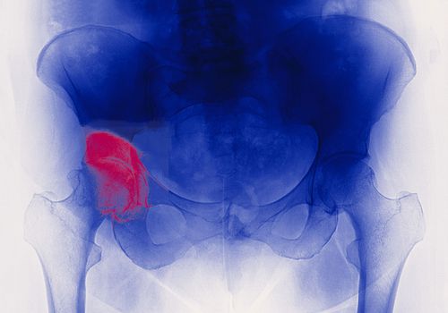
Femur neck fracture
Definition of femoral neck fracture
 The fracture of the neck of the femur is extremely common, especially in the elderly whose bones are weakened by osteoporosis. Most of the time this fracture is the consequence of a fall, but sometimes it is the spontaneous fracture of the porous bone that causes the fall. However, a young patient, during a major trauma, can also be a victim. The clinical signs are significant pain, the patient's inability to mobilize his lower limb, the lower limb is generally shortened and in external rotation. Pelvis and hip X-rays confirm the clinical diagnosis. The treatment is surgical. The choice of surgical technique depends on whether the fracture is located on the neck of the femur or on the upper end of the femur.
The different surgical procedures depending on the seat of the femur fracture
All the interventions below take place in the operating room in the orthopedic room under rigorously aseptic. The patient benefited from the usual skin preparation in the room before being taken to the operating room.
Osteosynthesis by screwing the neck of the femur
This surgical method is reserved for a small percentage of femoral neck fractures. The fracture is interlocked, the neck of the femur has impacted into the head of the femur, pain on bearing and mobilization raises suspicion of a neck fracture femoral. The patient can generally mobilize the lower limb. Some patients can walk despite the fracture, so it sometimes goes unnoticed for the first few days. Persistent pain leads patients to consult and only radiography can confirm the diagnosis. Without surgical intervention, the fracture risks moving secondarily and requiring more invasive surgery such as a hip prosthesis. The technique recommended in this type of fracture is a conservative intervention: osteosynthesis by screwing. The head and the neck are preserved, the screws come to maintain the fracture while waiting for the osseous consolidation. The patient is installed on a special operating table, "orthopedic table", which allows manipulation of the fractured limb throughout the intervention if necessary. After the usual skin preparation in the operating room, the sterile field is placed. Under radioscopic control the surgeon, after making a small incision on the external face of the upper thigh, places the screws in the neck and the head of the femur. If the quality of the bone is good, weight bearing is authorized postoperatively. The pain, which is not significant, allows a very rapid recovery of autonomy. If the quality of the bone is poor and causes fear of secondary displacement, weight bearing is prohibited or only partially authorized during the time of bone consolidation.
The hip prosthesis
In case of fracture of the neck of the femur moved, the indication of hip prosthesis is indisputable. During a high per trochanteric fracture, two alternatives present themselves, the hip prosthesis or osteosynthesis by intramedullary nail. The greater trochanter is the site of the insertion of many muscles. When the fracture preserves the area of muscle insertions or the greater trochanter can be reinserted, it is sometimes possible to fit a type of prosthesis designed specifically for these fractures. When the location of the fracture and that the general state of the patient allows it, the solution of the hip prosthesis is preferable the recovery of the autonomy being faster. Intermediate hip prosthesis means a prosthesis replacing only the femoral part of the joint. Apart from the first part of the operation aimed at replacing the acetabulum during a total hip prosthesis, the postoperative course and the complications are identical. See total hip prosthesis In disoriented patients, the risks of infection and prosthesis dislocation are increased because they do not demonstrate the necessary compliance in the postoperative period. This non-compliance with instructions and procedures increases postoperative risks.
Osteosynthesis by intramedullary nail
When the fracture is too low or the greater trochanter is fractured, the indication is then osteosynthesis with an intramedullary nail (gamma nail). The patient is installed on a special operating table, “orthopedic table”, which allows manipulation of the fractured limb and obtaining a good reduction for osteosynthesis. After the usual skin preparation in the operating room, the sterile field is placed. Through several incisions, under radioscopic control, the surgeon places the osteosynthesis material: the femoral nail, the cervical screw and the locking screw downstream. Weight bearing is authorized postoperatively depending on the stability of the osteosynthesis and the quality of the bone. If weight bearing is authorized, walking can be resumed the next day, however the pain sometimes lasts for several weeks, making the recovery of autonomy slower than after a hip prosthesis.
Risks and complications of femoral neck fractures
Phlebitis and its sequelae: despite the systematic implementation of anticoagulant treatment, phlebitis can form. Treated in time, phlebitis does not alter the functional results of the intervention. It is complicated by a pulmonary embolism only exceptionally.
The hematoma can form in the operating area and often resolves spontaneously, it rarely requires a gesture additional evacuation procedure.
The secondary displacement of the fracture
Surgical site infection