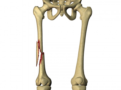
Femur fracture
Definition of Femur Fracture
 The fracture of the diaphysis of the femur generally occurs during a violent trauma, the femur being a resistant bone. The clinical signs are significant pain, the patient's inability to mobilize his lower limb, deformation (the thigh is shortened and increased in volume). Radiography confirms the clinical diagnosis. Treatment in adults is always surgical
Different surgical procedures
Depending on the site, the complexity and the type of fracture the therapeutic method will be different. All interventions below take place in the operating room in the orthopedic room under strictly aseptic conditions. The patient benefited from the usual skin preparation in the room before being taken to the operating room.
Long intramedullary nail osteosynthesis
The patient is installed on a special operating table “orthopedic table” which allows manipulation of the fractured limb and obtaining a good reduction for osteosynthesis. After the usual skin preparation in the operating room, the sterile field is placed. Through several incisions (external face at the top of the thigh and external face at the bottom of the thigh), under radioscopic control, the surgeon places the osteosynthesis material. The long femoral nail descending below the fracture, the cervical screw and the locking screw, upstream and downstream of the fracture. Weight bearing is prohibited postoperatively for two to three months, the time for bone consolidation. The patient can be lifted to the chair the next day to avoid complications related to bed rest. In a young patient, walking without support using a pair of crutches is acquired very quickly.
Osteosynthesis by intramedullary nail
The patient is installed on a special operating table “orthopedic table” which allows manipulation of the fractured limb and obtaining a good reduction for osteosynthesis. After the usual skin preparation in the operating room, the sterile drape is placed. The incision is made on the outer side of the thigh, long enough to visualize the entire fracture and place the osteosynthesis material. Once the reduction of the fracture obtained, the plate is placed against the femur and held in place by screws and possibly strapping Support is prohibited postoperatively for three months, the average time for bone consolidation. The patient can be lifted to the chair the next day to avoid complications related to bed rest. In a young patient, walking without support using a pair of English canes is acquired very quickly.
Retrograde nail osteosynthesis
The patient is seated on the operating table. After the usual skin preparation in the operating room, the sterile field is placed. Through several incisions (knee, external face and internal face of the thigh at its distal end), under radioscopic control, the surgeon places the osteosynthesis material. the retrograde nail going up above the fracture, the locking screws upstream and downstream of the fracture.