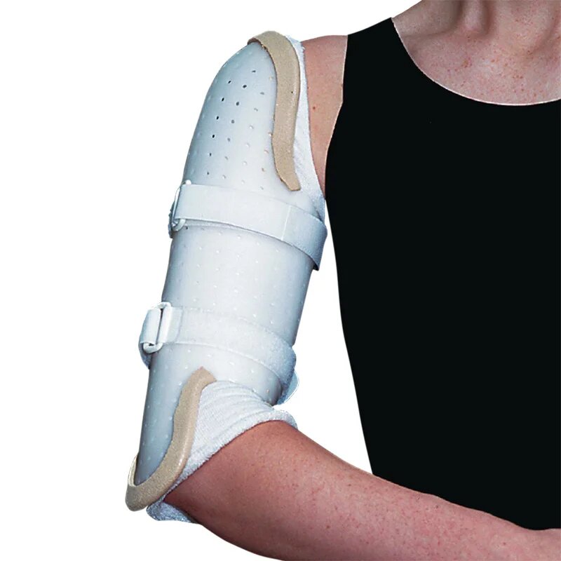
humeral diaphyseal fracture
Definition of shaft fractures of the humerus
 The clinical signs of humerus diaphyseal fractures are pain, swelling and inability to move the upper limb. X-rays confirm the clinical diagnosis. humerus is the lesion of the radial nerve, this nerve cleaving the bone in its middle third / lower third. The lesion may be caused by the fracture or by the edema caused by it.
Fracture of the humeral pallet
It is a serious fracture of the lower end of the humerus. In the event of a non-displaced fracture, the upper limb is immobilized for a month and a half. After bone consolidation, the immobilization is removed, rehabilitation can begin. The procedure takes place in the operating room under strictly sterile conditions. It can be done under general anesthesia, or locoregional anesthesia. The incision is made on the posterior side of the elbow, along the fracture to visualize it. After reducing the fracture, the surgeon places the osteosynthesis material using plates and screws or lost screws, sometimes using pins. The upper limb is immobilized with an elbow splint or a resin restraint for a month and a half. After consolidation, the immobilization is removed and rehabilitation to regain elbow mobility is started.
Orthopedic treatment
In the event of a non-displaced fracture, the upper limb is immobilized "elbow to the body" for three months . After bone consolidation, the immobilization is removed, rehabilitation can begin.
Surgical alternatives
All the interventions below take place in the operating room, under strictly aseptic conditions. The patient benefited from the usual skin preparation in the room before being taken to the operating room.
Osteosynthesis by intramedullary nail
This procedure is reserved for fractures of the upper two thirds of the humeral diaphysis. The patient is installed on the operating table. After the usual skin preparation in the operating room, the sterile drapes are placed. Through several incisions, under radioscopic control, the surgeon places the osteosynthesis material: the humeral nail which will be locked upstream and downstream of the fracture by screws. The upper limb is immobilized "elbow to the body" for a period that will depend on the stability of the assembly and then begins a gentle mobilization.
Retrograde pinout
The patient is installed on the operating table. operative, the sterile drapes are placed under radioscopic control, the pins are introduced via a mini approach above the elbow. The pins go up to the humeral head and aim to “roughly” align the fracture site. The upper limb is immobilized "elbow to the body" for three months.
Plate and screw osteosynthesis
The patient is installed on the operating table. operation, the sterile drapes are placed. The incision is made on the outer side of the arm, long enough to visualize the entire fracture and place the osteosynthesis material. The radial nerve is first identified and protected. Once the reduction of the fracture has been obtained, the plate is placed against the humerus and held in place by screws. The upper limb is immobilized "elbow to the body" for three months.