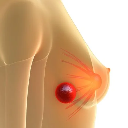
Breast nodule - mammary cancer
Definition
 Breast lumps are lesions of the breast tissue whose appearance can depend on various causes. Their presence may be accidentally felt by the patient during self-examination, or detected by the clinician during routine examinations (breast examination, breast ultrasound, and mammography).
The nodules may be painless or tender; sometimes they are accompanied by other signs, such as nipple discharge or skin changes.
Breast masses are a signal that should not be underestimated, but which should not cause undue concern: in 90% of cases, they are in fact benign nodular formations, such as fibroadenomas and cysts..
To remove doubts and differentiate between benign and malignant lesions, therefore to exclude the presence of a breast mass of neoplastic origin, it is always advisable to consult a specialist, who will prescribe a series of useful tests to identify its nature .
The management of breast masses depends on the causes and their histological characteristics.
Causes
The presence of a breast mass has many causes: it is often fibroadenoma, inflammation of all kinds or non-malignant fibrocystic alterations; Although very dreaded, the risk of a lump turning out to be breast cancer is very low.Certain benign nodular lesions may slightly increase the risk of developing cancer.
Fibrocystic mastopathy is the most common cause of breast lumps. It is a benign dysplasia (that is to say an abnormal development), quite common in women, especially between 30 and 50 years old. On palpation, these nodules are rounded and often appear as clumps in both sinuses or as well-defined, mobile masses with no signs of skin retraction. In fibrocystic mastopathy, nodular lesions enlarge and cause tenderness in the days before the onset of menstrual flow; the feeling of swelling and tension in the breast tends to disappear, then, at the end of menstruation.
Other fibrocystic changes that do not have neoplastic significance include adenosis (nodules of hard consistency and variable in size) and cysts (single or multiple round formations with liquid contents). Other nodules may be due to ductal ectasia and mild hyperplasia.
Fibroadenomas are benign solid nodules, typically painless and mobile (these lesions can be displaced under the skin of the fingertips), similar to small balls with sharp edges, circumscribed and receding. Usually, these lesions develop in young women (often in adolescents) and their characteristic mobility in the breast helps to distinguish them from other breast masses. A simple fibroadenoma does not appear to increase the risk of breast cancer, whereas a lesion with a complex structure may slightly increase the risk.
Breast infections ( mastitis ) cause severe pain, redness and swelling; an abscess resulting from this process can produce a lump that can be felt by touch. Mastitis is a rather rare condition and occurs mainly in the puerperal period (that is, in the postpartum period) or after a penetrating trauma. In addition, infections can appear after breast surgery. However, if an infection occurs in other circumstances, a tumoral origin must be quickly excluded.
Breast abscess is characterized by a painful mass that tends to gradually enlarge. The skin in the affected area is red, warm and "orange peel" in appearance. Sometimes the fever is associated with chills and general malaise. Breast abscess is more common during lactation and is a complication of mastitis.
In the postpartum phase, a galactocele may also appear, that is, a cyst rounded, movable and filled with milk. These cysts usually appear up to 6 to 10 months after the cessation of lactation and rarely become infected.
In addition to these etiologies, a breast mass can occur in the setting of tumors of various types. Breast cancer manifests as a hard, ill-defined lump that adheres to the skin or surrounding tissue. In this context, a deviation, retraction or flattening of the contour of the breast or nipple, with or without bloody or serous discharge, may also be evident. Other symptoms associated with breast cancer include redness and an "orange peel" appearance of the overlying skin, breast tenderness, and enlarged lymph nodes in the armpit (lymphadenopathy).
Signs and symptoms
Breast masses can be divided into benign lesions and malignant tumors. These formations are found on palpation or self-examination of the breasts and, in some cases, are visible to the naked eye.
Breast masses look like a kind of circumscribed peanut, with a different consistency from the rest of the breast, fixed or mobile.
Their presence can cause pain and may be accompanied by other signs, such as:
Leakage of fluid (serum or blood) from the nipple.
Skin changes (such as erythema and lymphedema with an "orange peel" appearance).
Feeling of tension.
Changes in breast shape or size.The presence of these manifestations could be the result of a scratch, inflammation or something else, to be studied again with the help of the doctor.
Benign lumps in the breast
Benign nodules have sharp outlines and are mobile, ovoid or rounded.
Depending on their nature, these lesions may tend to be solid (i.e. they have a hard consistency), fatty constitution (soft) or liquid content (cysts).
Malignant breast masses
Malignant nodules have ill-defined outlines (they tend to infiltrate the surrounding gland) and are not mobile. More advanced breast cancer almost always causes retraction of the overlying skin, with a change in breast shape and increased skin signs caused by lymphedema.
The presence of satellite nodules and lymphadenopathy is indicative of tumor propagation.
Potential Suspicious Signals
Among the symptoms that should make you suspect, and therefore should be reported to your doctor, are:
Sensation of one or more hard lumps in the chest or armpit.
Masses or thickening of the chest or armpits.
Changes in the areola of the breast or changes in the nipple (such as, for example, a clear milky discharge unusual or rash around the area).
Some signs are of particular concern:
Firm mass in the skin or chest wall.
Presence of nodular masses of very hard consistency, irregular in shape.
Integrated or fixed axillary lymph nodes.
Bloody discharge from nipple.
Appearance of dimpling or skin retractions, swelling, redness, heat and cracks.
Breast pain is however not a relevant symptom, as breast cancer remains indolent in most cases; however, it is best to talk to your doctor to reassure you.
Physical examination
Direct breast examination (breast exam) focuses on examining and feeling the breast and surrounding tissue. Feeling a mass by touch will reveal its size, tenderness, texture (i.e. hard or soft, smooth or bumpy) and mobility (whether it can be moved with the fingertips or stuck skin or chest wall).
During the evaluation, the mammary gland is inspected for changes in the area where the lump or mass is present, such as erythema, exaggeration of normal skin signs, lymphedema (skin of orange) and nipple discharge. The axillary, supraclavicular, and subclavicular regions are palpated for masses and lymphadenopathy.
Treatment
Treatment for breast lumps depends on the specific cause and may involve various therapeutic interventions.
Fibroadenomas can be excised by surgery under local anesthesia, but often recur.
To alleviate the symptoms of fibrocystic alterations, on the other hand, the use of analgesics (such as paracetamol) and the use of sports bras capable of providing adequate support and reducing transient soreness in the breasts may be helpful. In case of diagnostic doubt, surgical excision of the lesions may be indicated.
Usually, breast cysts do not require treatment unless the symptoms and size of these lesions are bother the patient. In these cases, it is useful to drain the fluid contained in the pocket-like formations by needle aspiration; although rarely, surgical removal may be indicated. After this procedure, the mammary gland is less tight and less painful, but breast cysts may re-form, as more fluid can accumulate inside.
In any case, breast masses should not be overlooked and their presence requires periodic monitoring by self-examination and ultrasound/mammography follow-up.