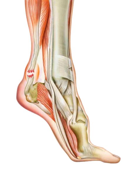
Achilles tendon rupture
Definition
 The rupture of the Achilles tendon occurs mainly during an intense muscular effort, the sudden contraction of the calf causing the rupture. The rupture of the Achilles tendon is accompanied by a tearing sound and a sharp but fleeting pain. The diagnosis is clinical, on palpation there is a depression in the tendon.
Orthopedic treatment
A resin immobilizer is made, keeping the foot in equinus (hyperextension) so as to bring the two parts of the tendon and let it heal. The patient should not press on the lower limb for three months and is given anticoagulant treatment during this period.
Lacing the tendon
The intervention takes place in the operating room under strictly aseptic conditions. The patient has benefited from the usual skin preparation in the room before being taken to the operating room. It can be done under general anesthesia or locoregional anesthesia. The surgeon and the operating room team will install you in a prone position on the operating table. After the usual skin preparation in the operating room, the sterile drapes are placed. The incision is made facing the tendon over approximately 8 cm, the surgeon suture the tendon and carefully refemre the operative wound. A splint is made on the front of the leg or a resin boot, maintaining the foot in equinus and avoiding constraints on the freshly sutured tendon. The patient should not press on the lower limb for 10 weeks, then the equine immobilization will be removed and he can resume weight-bearing. The patient receives anticoagulant treatment during this period. At the removal of the immobilization , physiotherapy will begin to regain good ankle mobility.
The tenolig
The procedure takes place in the operating room under strictly aseptic conditions. The patient benefited from the skin preparation for use in the room before being taken to the operating room. It can be done under general anesthesia or locoregional anesthesia. The surgeon and the operating room team install you in a prone position on the operating table. After the preparation cutaneous use in the operating room, sterile drapes are placed. Percutaneously, the surgeon passes two very solid threads from the muscular insertion of the tendon to its osseous insertion, the tensioning of these threads allows the confrontation of the two residues tendinous. The threads are attached to the skin on washers. A splint is made on the front of the leg, keeping the foot in equinus and avoiding stress on the tendon. The patient should not press on the lower limb for 10 weeks, then the equine immobilization will be removed and he can resume weight-bearing. The patient receives anticoagulant treatment during this period. The removal of these two threads is done after about 2 months.