Severe Traumatic Brain Injury (TBI)
Have you or a loved one suffered a severe traumatic brain injury? Neurosurgical management of severe traumatic brain injury in Tunisia is a specialized emergency management that allows effective treatment of post-traumatic brain injuries. Performed by experienced neurosurgeons, it offers high success rates at attractive prices.
What is severe traumatic brain injury?
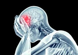 A severe traumatic brain injury (TBI) is a post-traumatic brain injury characterized by a Glasgow Coma Scale (GCS) ≤ 8 for at least 6 hours, or rapid neurological deterioration requiring emergency neurosurgical intervention. It is a life-threatening emergency involving functional prognosis (severe neurological and cognitive sequelae).
Traumatic brain injuries are classified into three categories according to the Glasgow Coma Scale. Mild TBI has a GCS of 14 to 15. Moderate TBI has a GCS of 9 to 13. Severe TBI has a GCS of 8 or less. Mild TBIs represent 90% of cases. Moderate TBIs represent 5% of cases. Severe TBIs represent 5% of cases.
Severe TBI is the leading cause of death and severe disability in young subjects (< 45 years). Mortality from TBI is 30 to 40% in adults, and the percentage of severe neurological sequelae (vegetative state, total dependence) reaches 20-30% of survivors. In Tunisia, our neurosurgery and neurosurgical intensive care services are equipped to manage these patients according to the recommendations of the Brain Trauma Foundation and the Société de Neurochirurgie de Langue Française (SNCLF).
What are the causes and associated injuries?
The main causes of severe traumatic brain injury are road traffic accidents. Falls from height are also a frequent cause. Domestic and sports accidents represent a part. Assaults are a less frequent cause. Brain injuries are classified as primary and secondary. Primary injuries are immediate such as contusions. Secondary injuries are early or late such as edema. Intracranial hypertension is a secondary complication. Post-traumatic cerebral ischemia is also a complication. Infections and post-traumatic epilepsy may occur.
What are the signs of severe traumatic brain injury? (Glasgow Coma Scale)
The Glasgow Coma Scale (GCS) is the standard neurological assessment tool. It evaluates three parameters:
| Parameter | Response | Score |
|---|---|---|
| Eye opening | Spontaneous | 4 |
| To voice | 3 | |
| To painful stimulation | 2 | |
| None | 1 | |
| Verbal response | Oriented | 5 |
| Confused | 4 | |
| Inappropriate words | 3 | |
| Incomprehensible sounds | 2 | |
| None | 1 | |
| Motor response | Obeys commands | 6 |
| Localizes pain | 5 | |
| Withdraws from pain | 4 | |
| Abnormal flexion (decortication) | 3 | |
| Abnormal extension (decerebration) | 2 | |
| None | 1 | |
| Total score | 3 to 15 | |
A Glasgow Coma Scale ≤ 8 defines severe traumatic brain injury.
How is severe traumatic brain injury managed in Tunisia?
Management follows the Brain Trauma Foundation (4th edition, 2016-2020) guidelines and the structured protocol for severe traumatic brain injury.
1. Pre-hospital management
Initial management of severe traumatic brain injury follows the ABCDE protocol. Intubation is performed in any patient with a GCS ≤ 8. Strict cervical spine immobilization is performed. Fluid resuscitation maintains mean arterial pressure ≥ 80 mmHg. The patient is transported to a center with 24/7 neurosurgery.
2. Emergency imaging workup
The imaging workup for severe traumatic brain injury is urgent. A non-contrast brain CT is performed in less than 15 minutes. A whole body CT is performed in case of polytrauma.
3. Neurosurgical treatment
Surgery for severe traumatic brain injury is specific to each lesion. An epidural hematoma is evacuated by craniotomy. An acute subdural hematoma is evacuated by craniotomy or craniectomy. Cerebral contusions are evacuated with or without craniectomy. A depressed skull fracture is elevated with dural repair.
4. Neurosurgical intensive care management (ICP monitoring)
Management of post-traumatic intracranial hypertension is rigorous. Target intracranial pressure is below 20-22 mmHg. Cerebral perfusion pressure must be above 60 mmHg. Treatment of refractory ICH uses hypertonic solutions and mannitol. Decompressive craniectomy may be performed. Barbiturates are also used.
Prognosis of severe traumatic brain injury
The prognosis of severe traumatic brain injury is variable. Overall mortality is 30 to 40% in adults. Favorable recovery is observed in 40 to 50% of survivors. Severe disability affects 15 to 25% of patients. Persistent vegetative state occurs in 5 to 10% of cases.
What is the price of management for severe traumatic brain injury in Tunisia?
Tunisia offers very competitive prices for the management of severe traumatic brain injury. Emergency brain CT costs between €200 and €400. Hematoma evacuation (subdural or extradural) costs between €3,000 and €6,000. Decompressive craniectomy costs from €4,500 to €8,000. Intracranial pressure (ICP) monitoring is included in the intensive care package. A complete package includes CT, surgery, hospitalization in neurosurgical intensive care (2 to 4 weeks) and start of rehabilitation. These prices are up to 60% cheaper than in Europe, without compromising the quality of care.
Frequently Asked Questions about Severe Traumatic Brain Injury in Tunisia
1What is severe traumatic brain injury?
Severe traumatic brain injury (TBI) is a post-traumatic brain injury with a Glasgow Coma Scale ≤ 8. It is a neurosurgical emergency involving vital and functional prognosis. Mortality is 30 to 40% and severe neurological sequelae occur in 20 to 30% of survivors.
2What are the signs of severe traumatic brain injury?
Signs include: prolonged loss of consciousness (> 30 minutes), Glasgow Coma Scale ≤ 8, pupillary abnormalities (anisocoria), signs of intracranial hypertension (bradycardia, hypertension), otorrhea, periorbital bruising (raccoon sign), post-traumatic seizures.
3How is TBI management carried out in Tunisia?
Management follows the guidelines of the Brain Trauma Foundation. It includes: intubation and ventilation, emergency brain CT, hematoma evacuation if necessary, intracranial pressure (ICP) monitoring and neurosurgical intensive care management.
4What is decompressive craniectomy?
Decompressive craniectomy involves removing a large bone flap to allow the edematous brain to expand, preventing cerebral herniation. It is indicated in case of refractory intracranial hypertension to medical treatments.
5What are the possible sequelae after a TBI?
Sequelae may include: hemiplegia, language disorders (aphasia), cognitive disorders (memory, attention), behavioral disorders, epilepsy, hydrocephalus, and in severe cases, persistent vegetative state.
6How long is the hospitalization for a TBI?
Hospitalization in neurosurgical intensive care generally lasts 2 to 4 weeks. A prolonged stay in a neurological rehabilitation unit of several months is often necessary for functional recovery.
7Is Tunisia equipped for TBI management?
Yes, Tunisia has modern neurosurgery and neurosurgical intensive care services, equipped with 128-256 slice CT scanners, ICP monitoring, dedicated neurosurgical operating rooms, and neurological rehabilitation centers.
Why choose Tunisia for the management of severe traumatic brain injury?
Tunisia has neurosurgeons and intensivists trained in the best European centers. Neurosurgery and intensive care equipment is modern. Intervention times are fast. ICP monitoring is systematic. All-inclusive packages include hospitalization, CT and surgery. Neurological rehabilitation is included. Management of chronic sequelae is available. Neurological rehabilitation centers allow prolonged stays.
In Conclusion
Severe traumatic brain injuries are neurosurgical emergencies that require rapid and specialized management. Tunisie Esthetic offers you quality care, performed by experienced neurosurgeons, in modern clinics, at attractive prices. Do not hesitate to contact us to discuss your situation and obtain a personalized quote.